Electron Paramagnetic Resonance (EPR) / Electron Spin Resonance (ESR)
Introduction to Electron Paramagnetic Resonance (EPR) and Electron Spin Resonance (ESR)
Electron paramagnetic resonance (EPR), also known as electron spin resonance (ESR) is a spectroscopic technique used to detect and characterize materials with unpaired electrons.
The principle of EPR relies on a static magnetic field that splits the spin-up and spin-down energy levels, called Zeeman splitting, as depicted in Figure 1a. In thermal equilibrium, there is a slightly higher population in the lower-energy Zeeman level, dependent on the strength of the magnetic field B and temperature. When the sample under investigation is irradiated with electromagnetic waves polarized orthogonal to the static field, transitions between adjacent Zeeman-split spin states can be induced if the radiation frequency matches the energy splitting E, according to:
Here, is the Landé g-factor for a free electron and denotes the Bohr magneton. Typically, the static magnetic field ranges from a few mT to 9 T resulting in microwave (MW) frequencies from 1 to 250 GHz (L- to G-band frequencies). Most commonly, EPR spectrometers operate near 9.5 GHz (X-band) and 35 GHz (Q-band)1.
The spin transition results in an absorption of MW energy, which can be recorded by a dedicated detector. Measuring the absorption as a function of magnetic field strength reveals the EPR spectrum of the sample under investigation, as shown in Figure 1b. In order to increase the signal-to-noise ratio of the weak absorption signals, the field modulation combined with lock-in detection yields a signal proportional to the first derivative of the absorption spectrum. Therefore, an additional pair of coils around the sample modulates the static magnetic field in a periodic manner (e.g., at 100 kHz), generating an oscillating measurement signal in the detector that is mixed with the driving signal and low-pass filtered by a lock-in amplifier. The lock-in amplifier output is therefore proportional to the first derivative of the absorption spectrum, resulting in the characteristic derivative-shaped EPR signal.
From the positions of the resonance peaks, researchers can reconstruct the spin Hamiltonian of the probed electron, including its magnetic coupling to surrounding electron and nuclear spins. Thus, by identifying the g-factor and hyperfine coupling constants of the Hamiltonian, they can obtain structural and dynamical information about the chemical bonding and elements in the nanoscale vicinity. Typically, organic free radicals, transition metal complexes, spin traps (organic molecules that trap the spin of a free radical and stabilize it), spin labels (paramagnetic molecules), and defects in solids can be investigated. From this data, EPR measurements provide unique insights into the identification of paramagnets, protein conformation, concentration quantification, monitoring redox reactions, and measuring their reduction potentials 2.

Figure 1: a) Zeeman splitting of the electron spin up and down energy levels dependent on the applied static magnetic field. b) Schematic showing a typical electron paramagnetic resonance absorption (top) and derivative (bottom) spectrum as a function of the magnetic field strength.
Synchronized Experiment Control in Pulsed Electron Paramagnetic Resonance
There are two main types of electron paramagnetic resonance (EPR) experiments: continuous-wave (CW) and pulsed EPR. In CW EPR the MW irradiation is applied throughout the whole measurement duration, while the static magnetic field is swept over a certain range, which includes the targeted resonance condition of the electron spins. In this way, the resonance peak in the measurement signal reveals the B-field strength of the resonance from which the g-factor can be calculated according to Eq. 1. For more sophisticated cases, there are also other parameters of the spin Hamiltonian that can be inferred. Although there are also more advanced schemes in CW EPR (such as ENDOR3), the amount of information gained is limited.
If the static field strength of the resonance condition is known at the given MW frequency (e.g., from a CW EPR measurement) and held constant, one can drive Rabi oscillations between the spin up and down states by applying MW pulses with a precise duration. By choosing the right pulse duration, one can rotate the spin state by 90° on the Bloch sphere (/2-pulse) or flip it completely by a -pulse.
Compared with CW EPR, pulsed EPR experiments provide more information about the complex local chemical environment of electron spins. This is because pulsed experiments enable the investigation of the electron spin’s relaxation mechanisms. Once the spin population is excited to a higher energy level by a pulse, the spin will spontaneously flip back to its lower-energy state, releasing the energy difference to the surrounding environment. This process is called spin-lattice relaxation and follows an exponential decay with a characteristic time constant
Applying a /2-pulse lets the spins precess with a defined phase on the equator of the Bloch sphere, imprinted by the phase of the MW pulse. This phase relation gets distorted due to spin-spin interactions with neighboring electron or nuclear spins. This leads to a decay of spin coherence characterized by the time constant .
Thus, measurements of the spins’ and/or relaxation times gives a much more precise picture of its local chemical environment and dynamics, similar to pulsed nuclear magnetic resonance (NMR) experiments. As an example, the spin coherence time can be measured by a Hahn-echo experiment, depicted in Figure 2, where a /2-pulse is followed by a -pulse after a variable delay time . During the first delay period, the spins dephase due to local magnetic field variations. The -pulse reverses the dephasing process, causing the spins to rephase after a second delay period , generating a measurable Hahn echo. As the delay time increases, the echo amplitude decays exponentially with the characteristic coherence time .
Despite this well-known Hahn-echo pulse sequence, there are more sophisticated sequences, such as pulsed electron-nuclear double resonance (ENDOR) or double electron-electron resonance (DEER), which use an MW and a radiofrequency field, or two MW fields, to probe a nuclear or electron spin coupled to the primary electron spin 1. As a technical prerequisite for these experiments, the experiment control unit has to deliver precise and synchronous pulses of variable duration to switch the MW source and gate the detector or data acquisition.

Figure 2. Hahn echo sequence pulse pattern with Bloch sphere spin evolution as well as measurement signal 4.
Common Challenges in pulsed EPR Experiments
The fast Rabi oscillation driven by the MW requires the experiment control to switch MW pulses with typical durations of 5-50 ns 1 with an even better precision. Therefore, performing high-quality pulsed EPR experiments involves overcoming several challenges, such as:
- Pulse Precision: Conventional electronics lack the resolution ( <1 ns) to define short, phase-controlled MW pulses needed for advanced pulse sequences.
- Phase control and flexibility: many sequences (e.g., Hahn-echo, DEER) need accurate phase cycling and variable delays. Complexity of multi-modal experiments: ENDOR, DEER, or ODMR experiments combine RF, MW, optical, and magnetic modulation, demanding precise coordination.
- Synchronization: Coordinating magnetic field sweeps, MW bursts, and detection requires tight timing (<100 ps jitter).
- Scalability: Advanced experiments (multi-pulse and multi-frequency EPR) require complex, programmable pulse schemes.
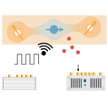
Swabian Instruments Pulse Streamer for Electron Paramagnetic Resonance Experiments
Swabian Instruments Pulse Streamer 8/2 digital pulse pattern generator provides a high-performance foundation for orchestrating ESR/EPR experiments:
- High timing precision: 1 ns timing grid with <50 ps RMS jitter allows for precise digital control of MW switches, detector gating, or triggering of data acquisition.
- Short pulse duration: 2 ns minimum pulse duration for the shortest MW pulses.
- Arbitrary pulse patterns: programmable digital pulse patterns on 8 synchronous channels effectively unlimited pulse sequence length via continuous streaming.
- High channel count: up to 14 digital and 4 analog channels by synchronizing 2 Pulse Streamer 8/2 systems.
- Analog channels for lock-in detection: 2 analog channels with 50 MHz bandwidth can provide the signals for the magnetic field modulation and reference input for the lock-in amplifier.
- Seamless software integration: application programming interfaces (API) for various languages like Python, MATLAB, or LabVIEW.
Summary & Conclusion
Since its birth in the 1940s 5, electron paramagnetic resonance (EPR) has emerged as a high-precision spectroscopy technique filling the gap between nuclear magnetic resonance and optical spectroscopy. Nowadays, it finds widespread application in chemistry, materials science, biology, and medicine. While CW EPR methods provide the spectrum of the sample under investigation, pulsed EPR experiments can reveal even more information by measuring the spin-lattice and spin-spin relaxation times.
However, pulsed EPR requires a precise yet fast timing control of the MW pulses, detector gating, and triggering of additional hardware. Swabian Instruments’ Pulse Streamer 8/2 perfectly meets these demands and orchestrates complex pulsed EPR measurements.
Relevant Resources
光探测磁共振 (ODMR)
Optically detected magnetic resonance (ODMR) is a powerful technique widely used in quantum sensing, magnetic field measurement, and material analysis.

Optically Detected Magnetic Resonance (ODMR)
Lindon JC, Tranter GE, Koppenaal DW, eds. Encyclopedia of Spectroscopy and Spectrometry. Third edition. Elsevier, Academic Press; 2017; pp. 527 - 534. ↩︎ ↩︎ ↩︎
Drescher M. EPR Spectroscopy: Applications in Chemistry and Biology. Springer Berlin / Heidelberg; 2012; p. 90. ↩︎
Weil JA. Electron Paramagnetic Resonance: Elementary Theory and Practical Applications. 2nd ed. Wiley-Interscience; 2007; p. 385. ↩︎
Spin echo animation by Gavin W. Morley, “GWM_HahnEchoDecay.gif”, Wikimedia Commons, GWM_HahnEchoDecay.gif, Licensed under CC BY-SA 3.0 Unported. ↩︎
Zavoisky E. Spin-magnetic resonance in paramagnetics. J Phys. 1945;(9):245. ↩︎